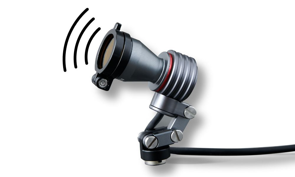
Today’s dermatoscopes are equipped with advanced optical and lighting systems designed to uncover what the naked eye can’t see, and one of the most useful of these is UV illumination, particularly at the 365 nm wavelength.
So, what makes UV light so special in dermatology, and why are clinicians turning to it for more precise skin analysis?
The Science Behind UV Illumination
At 365 nanometers, ultraviolet (UV-A) light interacts with the skin in a way that causes certain structures, compounds, and microorganisms to fluoresce, in other words, emit visible light when exposed to UV radiation.
This fluorescence helps reveal subsurface details that might otherwise go unnoticed under traditional white or polarized light. This helps get a clearer understanding of skin health, pigmentation, and potential pathology.
What UV Light Can Reveal
Pigment Disorders and Sun Damage: Under UV light, variations in melanin distribution become more visible, helping dermatologists and other clinicians distinguish between hypopigmented and hyperpigmented areas. This is helpful when evaluating vitiligo, melasma, and early sun damage that’s not yet visible under natural lighting.
Bacterial and Fungal Infections: Certain microorganisms naturally fluoresce at 365 nm:
-
-
Corynebacterium minutissimum (erythrasma) → coral-red glow
-
Pseudomonas aeruginosa → green
-
Malassezia (pityriasis versicolor) → yellowish-white
These visual cues make UV dermatoscopy a quick, non-invasive way to identify infections or confirm clinical suspicions.
-
Corynebacterium minutissimum (erythrasma) → coral-red glow
Porphyrins and Acne Activity: In acne-prone skin, UV light reveals orange-red fluorescence caused by porphyrins produced by Cutibacterium acnes. This helps practitioners assess inflammation, pore congestion, and the effectiveness of ongoing treatments.
Lesion Border and Structure Visualization: UV lighting can also enhance contrast in lesion morphology, making subtle edges, pigment networks, and vascular patterns more pronounced, a valuable aid in early melanoma detection.
Why 365 nm Is the Optimal Wavelength
Not all UV light is created equal. The 365 nm wavelength is considered ideal because it:
-
Maximizes fluorescence visibility for key skin chromophores and porphyrins
-
Produces minimal glare and skin reflection
- Penetrates safely without damaging the epidermis
Shorter UV wavelengths (below 320 nm) can be harmful, while longer ones (above 400 nm) lose diagnostic contrast, making 365 nm the “sweet spot” for clinical imaging.
A Dermatoscope Designed for Deeper Insight
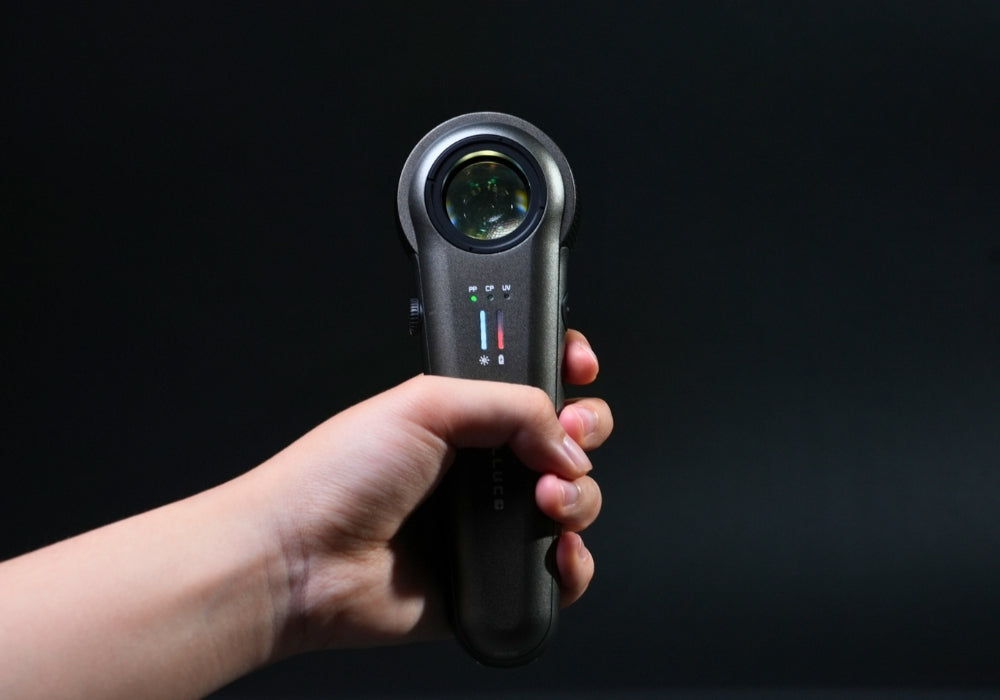
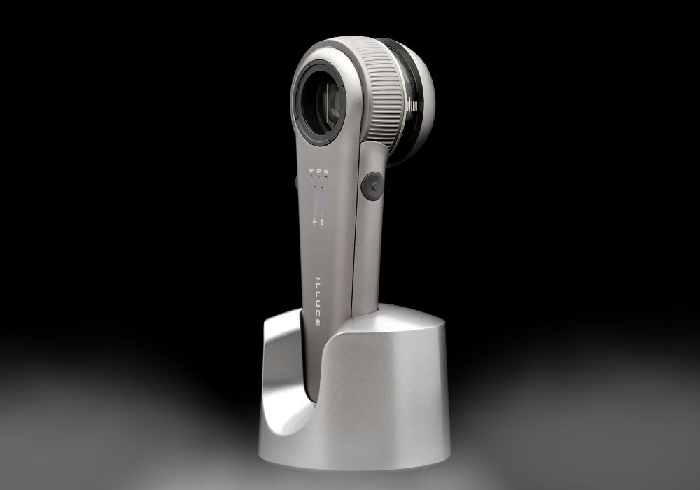
For clinicians seeking to integrate UV diagnostics seamlessly into their workflow, the ILLUCO IDS-9100 Dermatoscope provides this capability.
Featuring 12x optical magnification, it delivers exceptional clarity and color accuracy for detailed examinations. Its 12 light settings (3 modes × 4 brightness levels) include cross-polarized, parallel-polarized, and UV illumination at 365 nm, allowing for a tailored viewing experience across different skin conditions.
Built with a metal body, infection-control film, and a long-lasting rechargeable battery, the IDS-9100 is engineered for daily clinical use, providing the precision and durability professionals need without compromise.
UV illumination isn’t just a flashy feature; it’s a diagnostic tool that enhances your ability to see beyond the surface. Whether detecting subtle pigment changes, identifying infections, or assessing sun damage, 365 nm UV lighting adds an extra layer of clarity to your clinical evaluation.
With devices like the ILLUCO IDS-9100 Dermatoscope, this technology becomes an everyday part of practice, helping clinicians make more confident, informed assessments, one wavelength at a time.


Leave a comment
All comments are moderated before being published.
This site is protected by hCaptcha and the hCaptcha Privacy Policy and Terms of Service apply.