Frequently asked questions
Loupes FAQ
What is ILLUCO’s warranty on loupes and headlights?
Loupes: Oculars – 3 years, frames – 2 years
Headlights – 2 years, batteries – 1 year
Should I go for TTL or Flip-up loupes?
Choosing between dental loupes ultimately depends on personal preference. Each type offers its advantages.
Through-the-lens (TTL) loupes are lighter and less bulky, but they cannot be moved out of the line of sight when not in use and tend to be more expensive. They provide a wider field of view due to their close positioning to the eye, but peripheral vision is reduced, making reaching for instruments during treatment challenging.
Flip-up loupes, on the other hand, eliminate the need for customized fitting, reducing costs. They allow for easy adjustment of the declination angle, making them versatile for various procedures. Additionally, they can be moved out of the line of sight when not in use, offering added convenience. Plus, lending them to others is hassle-free since no customization is required.
Can I purchase headlights separately?
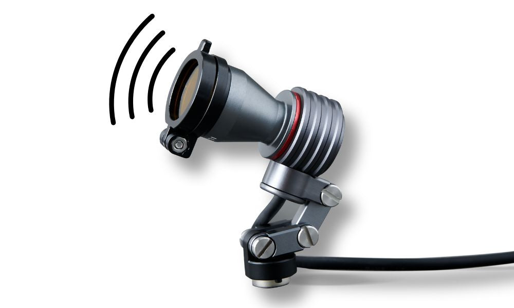
Certainly! You can buy headlights separately. We also offer non-prescription frames for those who only need headlights. Plus, we provide options like headbands or adapters to attach headlights to your glasses or loupes. You choose what works best for you!
Can I attach ILLUCO headlights to other brands’ loupes?
Yes, you can easily attach Illuco headlights to other brands’ loupes with a universal or clamp adapter. However, it should be noted that depending on the type of frame, attachment may be difficult, so this aspect needs to be considered.
Can the working distance be adjusted later?
Working distance is fixed at the time of purchase and cannot be adjusted afterward. If changes are required, adjustments can be made for an additional fee of $120. The product must be returned for modification and will be shipped back to you once the adjustments are complete.
Can I order the product if I have strabismus?
No, it's challenging to customize products for people with strabismus.
Can I take the composite filter off my headlight?
Yes. The composite filter can be unscrewed and removed from the light source.
Why are my loupes blurry?
If you're experiencing blurry vision when using your loupes, there could be a few reasons for it.
Firstly, incorrect adjustment or alignment might be the issue. Properly adjusting and aligning your loupes is crucial for clear vision. If they're not aligned correctly, the optics can be off, causing blurriness. It's a good idea to have the manufacturer help with adjusting them.
Secondly, if you wear corrective lenses, your loupes might need adjustments too. Sometimes, the magnification of the loupes can affect how well you see through your lenses. Consulting with an optometrist or the loupes manufacturer can help ensure your lenses and loupes work well together
Do I need a current Rx to order a new pair of loupes?
Yes, we will need a current (less than 2 years old) Rx to place the order. It is always good to get the most recent one.
Do you have a limitation on prescription (Rx) lenses?
Yes, the sum of SPH and CYL has to be lower than +10 or -10. All companies can only handle lenses at this range or lower.
Can I add my prescription (Rx) to the Flip-up Loupes?
While ILLUCO Flip-up Loupes are not custom-prescribed, they can be modified by your optician or optometrist after purchase to match your prescription.
Dermatoscopes FAQ
Which Dermatoscopes are polarized?
All of the devices have polarized light.
Which dermatoscopes can I take pictures with?
1100, 1100c, and 1000 Plus w/ phone camera accessories.
What's the lightest dermatoscope currently available?
1000/plus.
Which device offers UV light?
IDS3100.
Can I use it on my pets?
Yes, it is safe to use on pets.
How do I replace the battery for the IDS-3100?
The IDS-3100 uses a lithium-ion battery that is designed solely for the device. The replacement needs to be done by ILLUCO. Using a battery other than the one intended for the IDS-3100 can potentially cause damage to the unit and forfeiture your warranty.
How do I know my IDS-3100 is charging?
Three light indicators show the charging status of the IDS-3100:
- Red blinking: Low battery, charging required
- Green & Red: Charging
- Green: Fully charged
How long can I continuously use my IDS-3100?
Approximately 2.5-4 hours.
How do I clean and sanitize the device?
They can be cleaned and disinfected by isopropyl alcohol (70%). Warning, the use of disinfectant, especially acetone, may damage the device.
Note:
- Do not use any disinfectant, acetone, or chemicals other than isopropyl alcohol.
- Do not use alcohol directly on the lens; use it on a cloth or tissue.
- If there is a stain, first blow the dust off and apply alcohol on the stain.
- Important: Alcohol and Acetone are different chemical substances with different characteristics. Please make sure to distinguish them.
- To clean the lens, wipe using water or alcohol with the cleaning cloth with the device. There are alcohol-based cleaning fluids for lenses available on the market, which we recommend to have on hand. This will help remove fingerprints and other smudges without leaving streaks on your lens.
How do I clean the inside of the IDS-1000, IDS-1000 Plus, or IDS-1100 series?
When cleaning the internal part, never touch the LED & polarization filter. Clean the magnifying lens in the middle only if it has been contaminated. Use a lens cleaning cloth or a blower to remove the contaminations. Polarization filters are coated to protect themselves against foreign particles and dust as the filter itself easily attracts them like a magnetic field. If you clean the polarization filter using alcohol, fluids, or a cleaning cloth or tissue, the coating may peel off. Therefore, avoid wiping it. Instead, use a blower or air spray to remove foreign particles or dust if necessary.
How do I store my IDS-1000 or IDS-1000 Plus?
During work, permanently close the protective glass, keep the device with the protective glass side up, and store it safely. If you store IDS-1000/plus for a long time and the climate is very humid, use silica gel. In that case, do not use a leather pouch, as leather generally absorbs moisture in the air and causes mold. Do not store IDS- 1000/plus in a room with a temperature higher than 45°C/113°F
How do I replace the battery on the IDS-1000 or IDS-1000 Plus?
IDS-1000/plus is using a 2CR5 lithium battery. Please replace the battery when the indicator flashes red twice. After blinking red, the device will turn off automatically.
How to do Non-Contact Examination?
You can remove the Protect Glass for non-contact examination. The magnetically coupled Protect Glass (PG) can be easily removed using your fingernail.
What kind of battery can be used with the IDS 1100/C?
IDS-1100 uses a lithium-ion battery designed solely for ILLUCO IDS-1100. Please only use lithium-ion batteries from ILLUCO or an authorized ILLUCO dealer. Using a battery other than the one intended for the IDS-1100 can damage the unit.
How do I clean the protective glass of my device?
The scale on the protective glass is chrome-plated, so you can clean it with alcohol without worrying about being wiped off. A dermatoscope is classified as a non-critical instrument. Therefore, low-level disinfection methods are requested. Use 70% Ethanol or Isopropanol for disinfection. Non-contact examinations are asked when scoping damaged or infected skin. Even so, regular exhaustive disinfection is necessary, and the EO (Ethylene oxide) Gas sterilization method is recommended for deep disinfection.
How do I store the IDS-1100/C?
Use silica gel if you store IDS-1100 for a long time and the climate is humid. In that case, do not use a leather pouch, as leather generally absorbs moisture in the air and causes mold. Do not store IDS-1100 in a room with a temperature higher than 45℃/113°F
How do I troubleshoot the IDS-1100/C?
The IDS-1100 is reliable and designed for ease of use. Never attempt to open or dismantle the device for any reason other than battery replacement. The manufacturer and distributors are not liable for mechanical troubles, property damage, or personal injury caused by users (s) unfamiliar with the manual's instructions.
My device won't turn on. What should I do?
Recharge the battery and power it on again. If the problem persists, replace the battery with a new one. If the problem persists, please contact the ILLUCO A/S Center or your local distributor and report the problem.
My device is not charging. What should I do?
Change the charging cable. If the problem persists, replace the battery with a new one. If the problem persists, please contact the ILLUCO A/S Center or your local distributor and report the problem.
What should I do if an LED light is not working?
The LEDs embedded in IDS-1100 are designed to last over 100,000 hours. If any of the LEDs fail, there can be cracks in the soldering. In that case, contact the ILLUCO A/S Center or your local distributor and report the problem. If there are issues with functional buttons and focusing wheels, please get in touch with the ILLUCO A/S center or your local distributor.
Can I fix my device if it malfunctions?
If you attempt to disassemble it, there is a high probability of breakage on connections and joints and cause the warranty to be voided.
Dermoscopy FAQ
Why is polarization necessary when performing dermoscopy?
Polarization is essential when performing dermoscopy because it gives us a more in depth look/visualization of the deeper structures in the skin, such as blood vessels and collagen, which are not as visible with nonpolarized dermatoscopes. Polarized light can penetrate deeper into the skin and capture backscattered light from the dermal layers, enhancing the visualization of the DEJ and dermal structures. Nonpolarized dermatoscopes, such as milia-like cysts, are better for visualizing superficial structures in the epidermis. Familiarity with nonpolarized and polarized dermoscopy is crucial for understanding the pertinent applications of each modality in evaluating pigmented and non-pigmented skin lesions.
Is dermoscopy possible without polarization?
Yes, dermoscopy is possible without polarization. Non-polarized dermoscopy is another modality of dermoscopy that provides additional information beyond that gleaned by evaluating the lesion through a simple magnifying lens. It requires a liquid interface and direct contact with the skin. For most pigmented and non-pigmented skin lesions, polarized and non-polarized dermoscopies offer similar images. However, some dermatoscopes provide higher-quality visualization of dermoscopic structures and colors when used with immersion fluid and direct skin contact. Hybrid dermatoscopes have also been developed that allow the user to toggle between polarized and nonpolarized modes, but they should be applied using direct skin contact with a liquid interface. If this is not done, then the user will see dermoscopic structures only in the polarized mode, and no dermoscopic systems will be discernible in the non-polarized mode; instead, the observer will see a magnified clinical (not dermoscopic) image of the surface of the lesion.
Is liquid always necessary when performing contact dermoscopy?
No, liquid is not always needed when performing contact dermoscopy. While some dermatoscopes require immersion fluid and direct skin contact for higher-quality visualization of dermoscopic structures and colors, other devices, such as those using cross-polarization, do not mandate the use of a liquid interface and do not require direct contact with the skin. However, it is essential to note that PD typically requires more vital LED lighting to compensate for the photons blocked by cross-polarization.
Is dermoscopy only helpful for the evaluation of pigmented lesions?
No, dermoscopy is beneficial in assessing both pigmented and non-pigmented skin lesions. Significant progress has been made in defining benign and malignant dermoscopic structures and patterns of both lesions, making dermoscopy a valuable clinical tool for the noninvasive, in vivo evaluation and diagnosis of cutaneous lesions. Familiarity with non-polarized and polarized dermoscopy is crucial to understanding pertinent applications of each modality in evaluating pigmented and non-pigmented skin lesions. Dermoscopy can also aid in the detection of pigmented structures that are not visible to the naked eye, helping to select appropriate treatments for non-pigmented basal cell carcinomas (BCCs) and detect residual/recurrent BCC after noninvasive therapies.
What's the typical magnification used in dermoscopy?
Dermoscopy can be performed using a handheld dermatoscope with 10-fold magnification. However, digital dermatoscopes ranging from 20 to 1000-fold magnification have the additional benefit of allowing more precise measurements of the visualized structures, making them suitable for monitoring disease severity.
To what degree does dermoscopy enhance the accuracy of diagnosis?
Dermoscopy significantly improves the specificity and sensitivity for detecting skin cancer, including melanoma. Studies have shown that dermoscopy alone can result in potential diagnostic pitfalls, so a more integrative approach to diagnosis is recommended to improve diagnostic accuracy further. Sensitivity measures the proportion of melanomas correctly diagnosed as suspicious and surgically removed. In contrast, specificity measures the proportion of benign lesions correctly diagnosed as benign and spared from unnecessary biopsies. Dermoscopy combined with sequential digital dermoscopy imaging (SDDI) has been shown to double the sensitivity for melanoma and reduce the excision or referral rate of benign lesions by more than 50%. Using both techniques in a "multimodal approach" can increase melanoma diagnostic accuracy. Knowledge of histopathological correlates can also be helpful in accurately interpreting clinical and dermoscopic information.
Does insurance generally reimburse for dermoscopy?
Insurance coverage for dermoscopy varies depending on the specific insurance plan and provider. Some insurance plans may cover dermoscopy as a diagnostic tool for skin cancer screening, while others may not. It is recommended to check with your insurance provider to determine if dermoscopy is covered under your plan. Additionally, some dermatology clinics may offer dermoscopy as a self-pay service for patients without insurance coverage.
How is UV (Woods) lighting beneficial when performing skin exams?
- Fluorescence: Certain substances, such as skin cells infected with fungi or bacteria, will fluoresce when exposed to UV light. This means they will emit light of a different color than the UV light on them. This can help doctors to identify and diagnose skin infections.
- Differentiation of skin types: UV light can also differentiate between skin types. For example, fair-skinned people will have a more pronounced red or pink fluorescence than dark-skinned people. This can be helpful for doctors when making diagnoses and determining the best course of treatment. IDS 3100 offers UV lighting
Which dermoscopic signs are most specific for melanoma?
Asymmetry in the distribution of colors and structures within a lesion is considered the best predictor of malignancy, followed by blue-white structures and atypical networks. Lesions with a score of two or three points are considered positive, and a skin biopsy or referral is recommended. The three-point checklist has a sensitivity of 79% to 91% and a specificity of 71–72% for diagnosing melanoma and BCC. It is recommended that any pigmented lesion with focal adherent keratin or a rough texture that reveals an asymmetric dermoscopic pattern be considered suspicious to avoid missing the diagnosis of malignancy.
Can you describe the 3-point checklist?
The 3-point checklist is a skin cancer screening tool with high sensitivity for pigmented skin cancers, including pigmented BCC and melanoma. It includes three dermoscopic features: asymmetry of pattern and structures, blue-white structures, and atypical network. Asymmetry is an asymmetry in the distribution of dermoscopic color and structures in one or two perpendicular axes. Blue-white structures are defined as a blue-white veil, white scar-like depigmentation, and blue pepper-like granules. An atypical network is a pigment network with thick lines and irregular-sized holes.
What are some of the more common mistakes made when performing dermoscopy?
Some common mistakes made when performing dermoscopy include false positive and false negative diagnoses, which can result in unnecessary excisions of benign lesions or delays in detecting malignant tumors. Collision tumors, featureless lesions, and lesions presenting with structures characteristic of other diagnoses are also prone to misdiagnosis. It is essential for dermoscopists to correctly assign weight to clues and avoid overvaluing unreliable or misleading indications. Experience and training are necessary for mastering dermoscopy.
Can dermoscopy assist in the diagnosis of actinic keratoses?
Dermoscopy cannot always reliably differentiate pigmented actinic keratosis from lentigo maligna, but it may help guide the best location for biopsy. Biopsying areas revealing the most suspicious features, such as annular-granular structures, asymmetric follicular openings, dots within the ostial spaces, or rhomboidal structures, may provide the pathologist with the most diagnostically relevant tissue to examine. However, caution should be exercised when not performing a complete biopsy of any lesion to exclude or confirm melanoma.
How does squamous cell carcinoma present in dermoscopy?
Squamous cell carcinoma can present in dermoscopy with atypical network and regression structures. Invasive SCC arising in pigmented Bowen's disease may show dermoscopic features of the latter, such as a dermoscopic blue and white structureless area and white circles. When evaluating non-pigmented tumors, dermatologists should consider clinical features, dermoscopic keratin clues, dermoscopic morphology of vascular structures, architectural arrangement and distribution of vessels seen under dermatoscopy, and additional dermoscopic criteria such as the rosette sign or strawberry pattern. Experienced dermoscopists can usually diagnose in a few seconds, as most skin neoplasms exhibit repetitive morphologic characteristics that are easily recalled and recognized. It is recommended to perform a total body skin examination on patients with a history of skin cancer, patients with a family history of melanoma, patients under the age of 50 years who present with more than 20 nevi on the arms, and patients over the age of 50 years who present with evidence of chronic solar damage.

